Imaging-derived Extranodal Extension (iENE) in Head and Neck Cancer

A Guide for Radiologists from the AOSHNHR-ASHNR-ESHNR Joint Task Force on iENE

Round 3: Complex Cases and Borderline Cases

Grade 0

Grade 1

Grade 2

Grade 3

Grade 0 (not Grade 3)

The border between the node (arrow) and the submandibular gland (asterisk) is ill-defined. But it appears that the gland is draped over (displaced by) the node, rather than definitively invaded. This scan might be reported as “probable iENE”, but it is not sufficient evidence for Grade 3 and upclassifying the neck nodes.

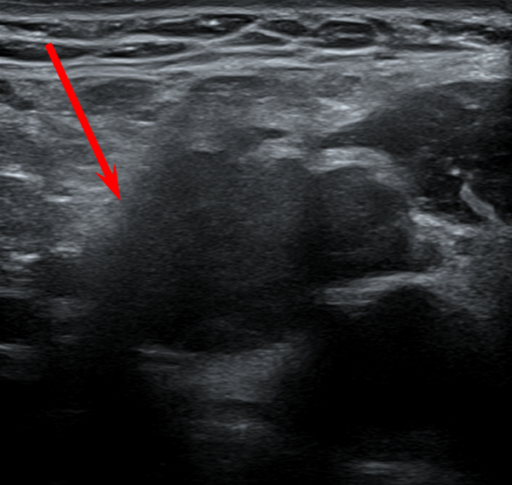
Grade 0

The margin of the node is not well-defined, which might prompt a diagnosis of Grade 1 iENE. But there is artifact in this area and the findings are not definitive, so Grade 0 is most appropriate.

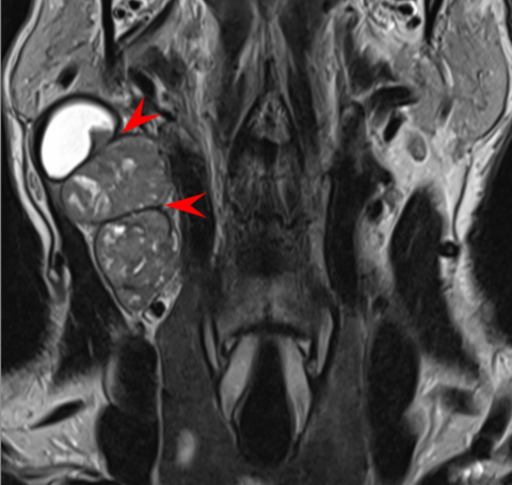
Grade 0

Coronal T2 images suggest Grade 2 iENE because the nodes are adjacent. But acute angles between nodes are preserved, so the three criteria for Grade 2 iENE are not met. Be sure to use multiple imaging planes to establish any Grade of iENE (but especially Grade 2).

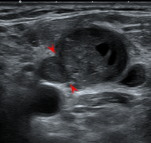
Grade 0

The fat planes between these nodes have been lost, but the acute angles at the edges of the contact planes are preserved (arrowheads). This is probable coalescence, but definitive coalescence is needed for establishing Grade 2 iENE. (The contralateral skin lesion is unrelated.)

Grade 1

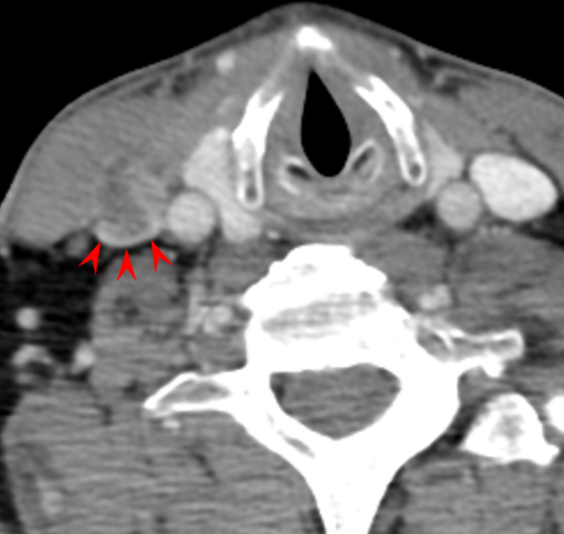
There is extensive invasion of surrounding fat planes, rendering this node Grade 1 iENE. Strands of enhancement extend into the SCM (arrow). This is likely invasion of muscle, but it is not sufficient evidence to conclude definitive Grade 3.

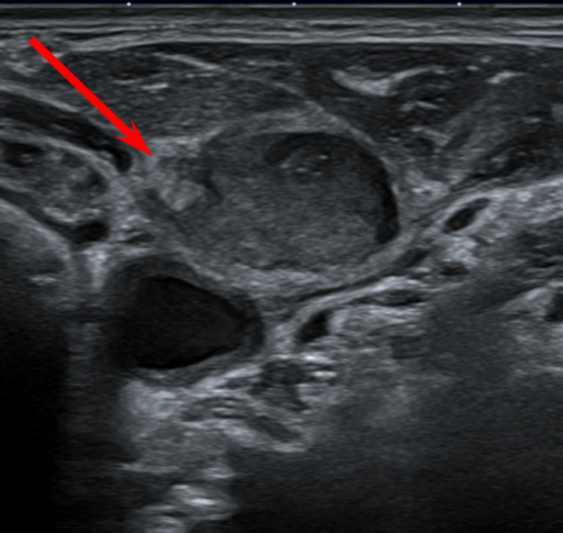
Grade 1

The margins of this node are irregular and ill-defined. The extension into surrounding fat is not uniform, with more invasion anterior and posterior (arrow). Therefore, this should be considered definitive Grade 1 iENE.

Grade 1

There are finger-like projections of tumor along the border of this node. This configuration is sufficient to establish Grade 1 iENE.

Grade 2

Numerous nodes have become coalescent (arrow). The nodes abut the SCM and paraspinal muscles but there is no DEFINITIVE invasion, so Grade 3 should not be applied.

Grade 2

All three imaging criteria for coalescent nodes are met:

  1. loss of fat plane between the nodes
  2. loss of convex border of the nodes
  3. loss of acute angle between the nodes (arrowhead)

Grade 2

Although some lobulation can be expected within a single pathologic node, the lobulation in this node is sufficient to merit a designation of definitive Grade 2 iENE, once confirmed on multiple images in multiple planes.

Grade 2

Although some lobulation can be expected within a single pathologic node, the discrete line differentiating these two coalescent nodes indicates that it is not a single node with lobulation. Because it meets all three criteria for coalescent nodes, this should be interpreted as Grade 2.

Grade 2

Three left-sided nodes are depicted, with confluent enhancement. The central node is cystic and the others are solid. Contrast this with the previous example, where a single node was mixed cystic-and-solid. The coalescent nodes are sufficient for Grade 2 iENE.

Although the nodes abut the overlying SCM, the signal intensity within the muscle remains normal, so muscular invasion is NOT definitive.

Grade 3

The jugular vein is often displaced or compressed by adjacent nodes, which would not indicate iENE. In this case, however, there is definitive invasion of the vein with a claw sign around the posterior margin (arrowheads) and tumor thrombus within the vein. Similarly, if the vein were completely obliterated, that would indicate Grade 3 iENE. Muscle, glands, skin and neurovascular structures all fall under the definition of “surrounding organs”.

(The main bulk of the node is more superior and is not shown on this image.)

Grade 3

There is an extensive nodal coalescence, which would be sufficient for Grade 2, but there is also abnormal signal within the SCM (arrow), which indicates tumor invasion of muscle. To better appreciate the signal change within the SCM, compare it to its counterpart on the other side of the neck. The findings of Grade 3 iENE supersede the Grade 2 findings.

Grade 3

All three Grades of iENE are depicted on this image. The dominant finding is the infiltration of the fat around the lateral node (Grade 1). The lateral node is coalescent with the medial nodes in the cluster (Grade 2). The nodal mass erodes through the SCM and platysma muscles to reach the skin surface (Grade 3). When multiple Grades of iENE are present, the patient receives the highest Grade (in this case, Grade 3).

Grade 3

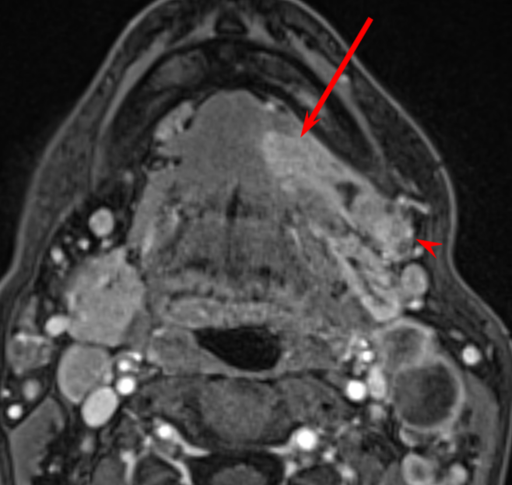
The oral cavity primary tumor arising in the floor of mouth (arrow) has become confluent with a submandibular lymph node (arrowhead). This may also happen with primary hypopharyngeal tumor extending into nodes in the internal jugular chain (Level III).

This is considered Grade 3 because of the invasion of intervening muscle structures such as the mylohyoid muscle or pharyngolaryngeal muscles.

Nasopharyngeal carcinoma that is confluent with retropharyngeal nodes is a special case because RP nodes are not considered when grading iENE in NPC!

Grade 3

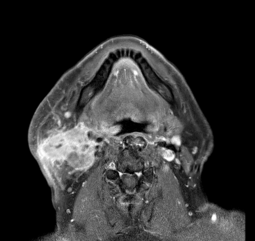
The T2-weighted image is not convincing because of the low-intensity rim around the lymph node that appears to separate it from the nearby soft tissues. But the post-contrast image shows definite invasion around the anterior aspect of the SCM, as well as the parotid gland. Remember to use all the sequences; don’t rely on just a single preferred sequence.

Grade 3

Retropharyngeal nodes (from a non-NPC primary) extending into the longus capitus muscle and encasing the internal carotid artery is enough to call this Grade 3.  But also note spread into the hypoglossal canal (arrow) which may be caused by tumor spreading along a path of least resistance or may be caused by true perineural spread. Secondary findings of muscle denervation in the tongue from a 12th nerve palsy would make this case definitive Grade 3 based on nerve invasion, even without the arterial and muscular findings.