
- 04/24/2025
- Posted by: ASHNR
- Category: Case of the Week
No Comments
Click the arrow to see the next slide with the correct interpretation.


One Month FU Sub-cm Lytic Lesion. Normal Labs. Biopsy: Lymphatic and Vascular Tissue. No Malignancy or Infection.
This radiologic case highlights a sub-centimeter lytic lesion discovered during a follow-up (FU) scan. Despite its worrisome appearance, lab results were normal and biopsy revealed only lymphatic and vascular tissue, ruling out malignancy or infection.
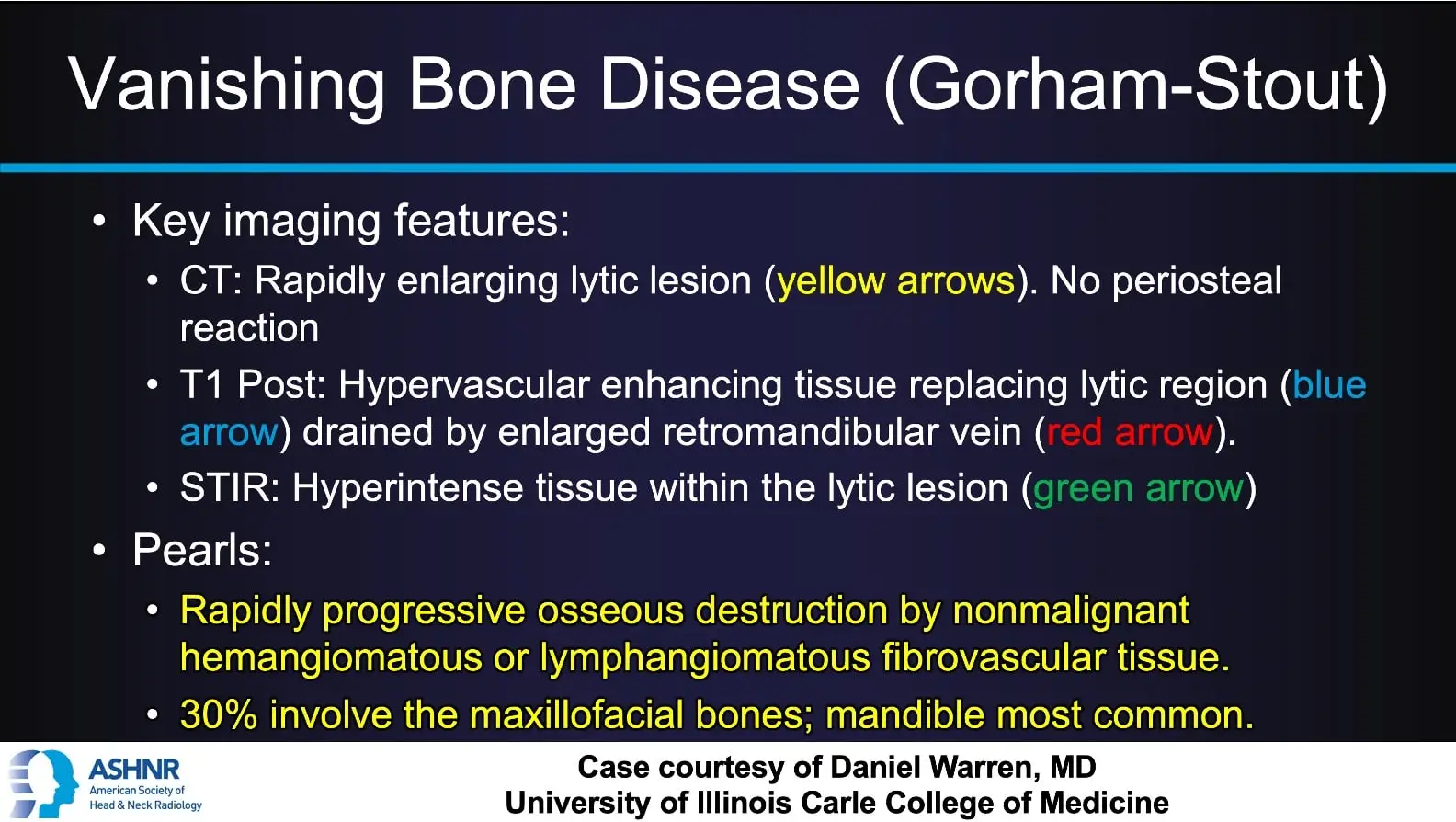
The diagnosis was Vanishing Bone Disease (Gorham-Stout), a rare condition characterized by progressive bone loss replaced by benign vascular tissue.
Key Findings:
- CT (Axial CT Face): Shows the lesion with no periosteal reaction, marked by yellow arrows.
- MRI T1 FS Post-contrast: Reveals hypervascular tissue replacing the bone, with drainage via an enlarged retromandibular vein (blue and red arrows).
- MRI STIR: Demonstrates hyperintense signal within the lesion (green arrow), consistent with fluid-rich or vascular tissue.
The condition commonly affects the mandible and maxillofacial bones and is caused by nonmalignant hemangiomatous or lymphangiomatous tissue.
Case courtesy of Daniel Warren, MD
University of Illinois Carle College of Medicine