

Case of the Week
-
Case 355
- 09/18/2026
- Posted by: ASHNR
- Category: Case of the Week
No Comments
Left visual disturbance on MRI shows an enhancing orbital mass with reduced diffusion. Learn why this points to orbital lymphoma.
-
Case 354
- 09/11/2026
- Posted by: ASHNR
- Category: Case of the Week
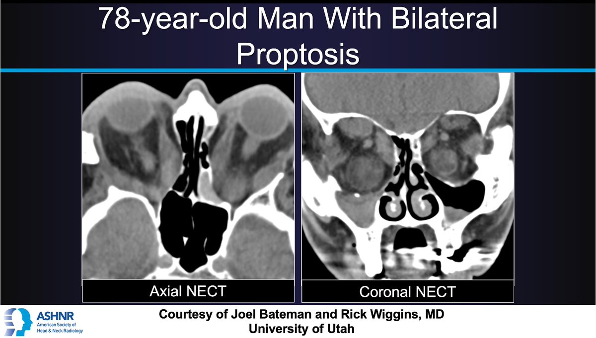
Bilateral proptosis in a 78-year-old man on CT shows enlarged, fatty extraocular muscles. Learn why this points to thyroid eye disease.
-
Case 353
- 09/04/2026
- Posted by: ASHNR
- Category: Case of the Week

Sinus CT in silent sinus syndrome presenting as orbital asymmetry, with maxillary atelectasis and enophthalmos. Case by Chen and Warren, MD, Carle Foundation Hospital.
-
Case 352
- 08/23/2026
- Posted by: ASHNR
- Category: Case of the Week
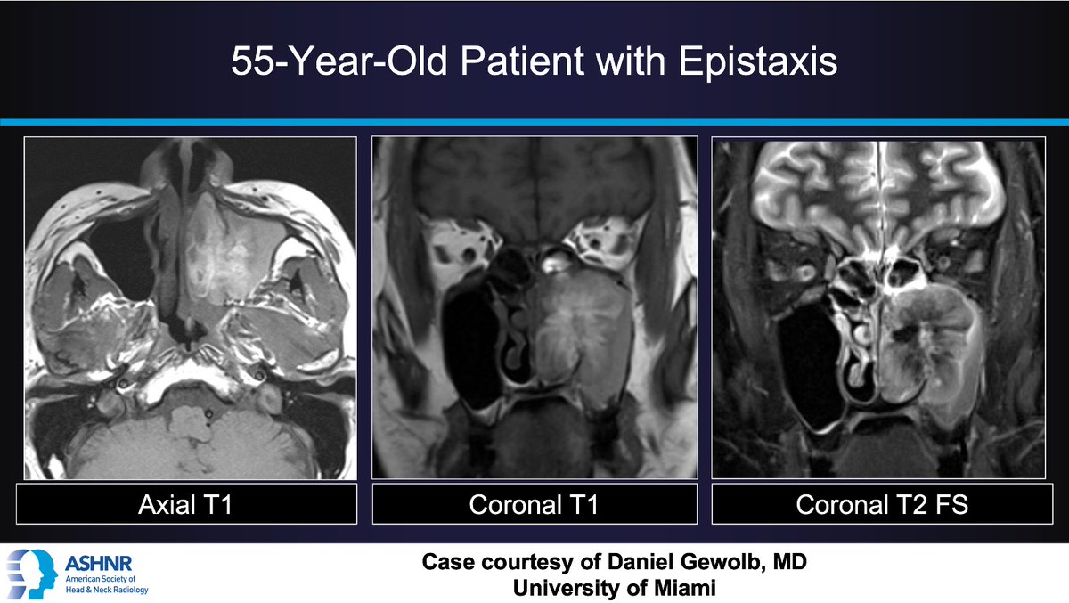
Sinonasal MRI in sinonasal melanoma presenting as epistaxis in a 55-year-old, with T1 hyperintensity and septate pattern. Case by Daniel Gewolb, MD, University of Miami.
-
Case 351
- 08/14/2026
- Posted by: ASHNR
- Category: Case of the Week

Neck CT in Mycobacterium avium cervical lymphadenitis presenting as persistent neck swelling in a 3-year-old despite antibiotics. Case by Alexandra Foust, DO, Vanderbilt.
-
Case 350
- 08/10/2026
- Posted by: ASHNR
- Category: Case of the Week
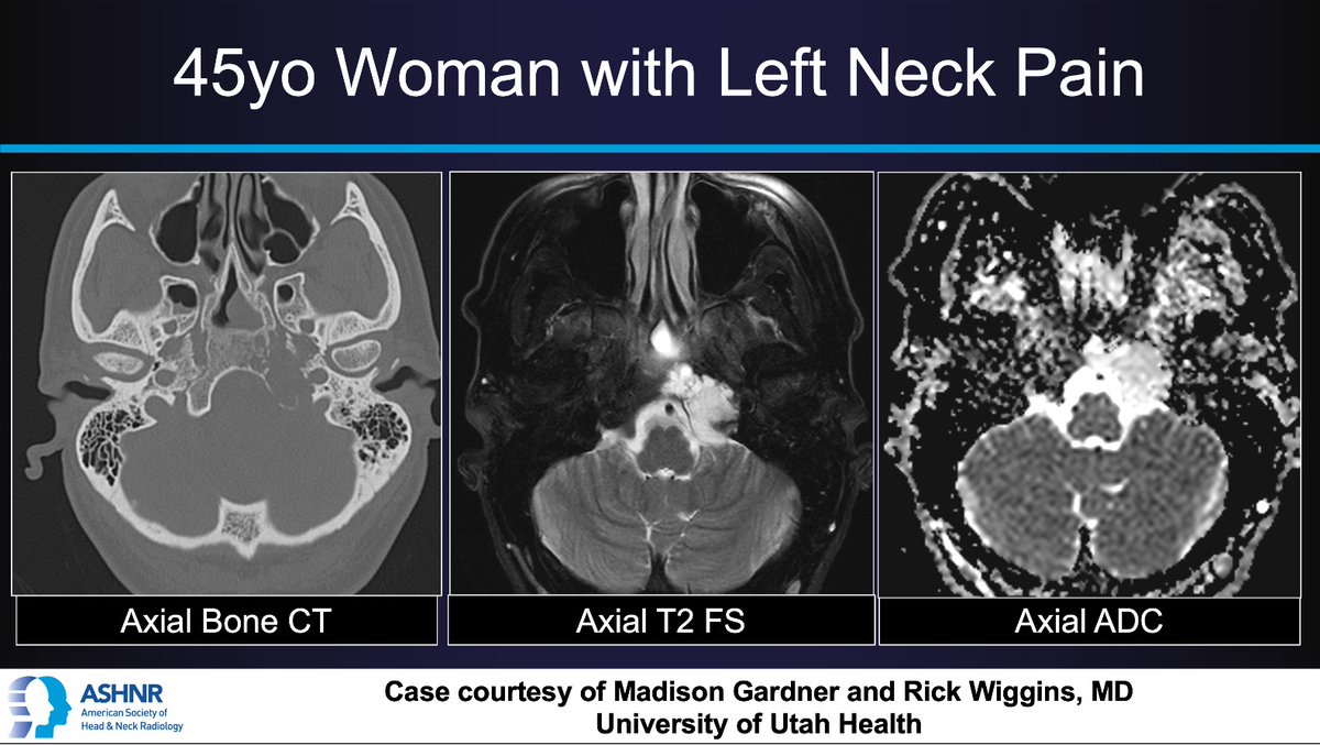
Skull base CT and MRI in chondrosarcoma presenting as left neck pain in a 45-year-old woman, centered at the petro-occipital suture. Case by Gardner and Wiggins, MD, Utah.
-
Case 349
- 07/31/2026
- Posted by: ASHNR
- Category: Case of the Week

Cheek MRI in minor salivary gland mucoepidermoid carcinoma presenting as an enlarging left cheek mass in a 29-year-old male. Case by Immen and Vartevan, DO, Camelback Radiology.
-
Case 348
- 07/24/2026
- Posted by: ASHNR
- Category: Case of the Week
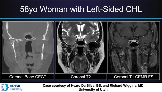
CT and MRI in TMJ pigmented villonodular synovitis presenting as conductive hearing loss in a 58-year-old woman. Case by Da Silva and Wiggins, MD, University of Utah.
-
Case 347
- 07/17/2026
- Posted by: ASHNR
- Category: Case of the Week
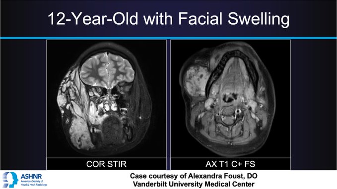
Facial MRI in venous malformation presenting as facial swelling in a 12-year-old, with T2 hyperintensity and puddling enhancement. Case by Alexandra Foust, DO, Vanderbilt.
-
Case 346
- 07/10/2026
- Posted by: ASHNR
- Category: Case of the Week
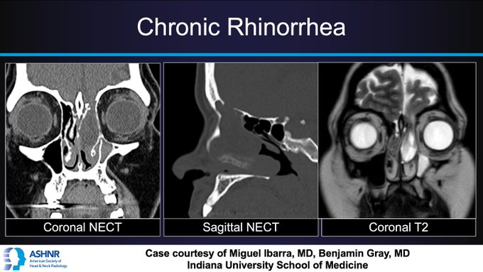
CT and MRI in meningoencephalocele presenting as chronic rhinorrhea, with fovea ethmoidalis defect and brain herniation. Case by Ibarra and Gray, MD, Indiana University.