
Click the arrow to see the next slide with the correct interpretation.


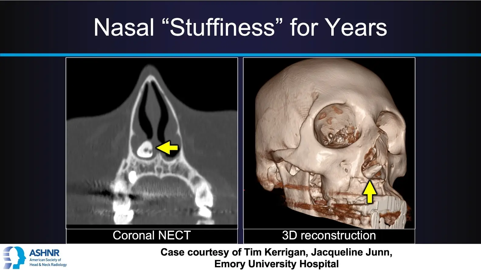
Nasal “Stuffiness” for Years
This case features a patient with long-standing nasal obstruction, ultimately diagnosed with an ectopic supernumerary tooth in the nasal cavity.
Imaging overview:
Coronal NECT and 3D CT reconstruction show a tooth-density structure along the floor of the nasal cavity, with a hyperdense rim and hypodense center corresponding to the pulp cavity. Surrounding soft tissue thickening suggests granulation tissue. CT clearly defines the relationship of the ectopic tooth to the nasal floor and adjacent structures, guiding surgical planning.
Clinical insight:
Ectopic or supernumerary teeth are uncommon but can occur in the nasal cavity, where they may present as chronic unilateral “stuffiness,” obstruction, crusting, or epistaxis. On imaging, they match the density of teeth elsewhere in the maxilla and may be seen clinically as a white mass surrounded by inflamed or granulation tissue. Recognition of this characteristic appearance helps avoid misdiagnosis as a foreign body or tumor and supports timely ENT referral for endoscopic removal.
Case courtesy of Tim Kerrigan and Jacqueline Junn
Emory University Hospital.