
Click the arrow to see the next slide with the correct interpretation.


Skull Base Lesion in a Young Adult
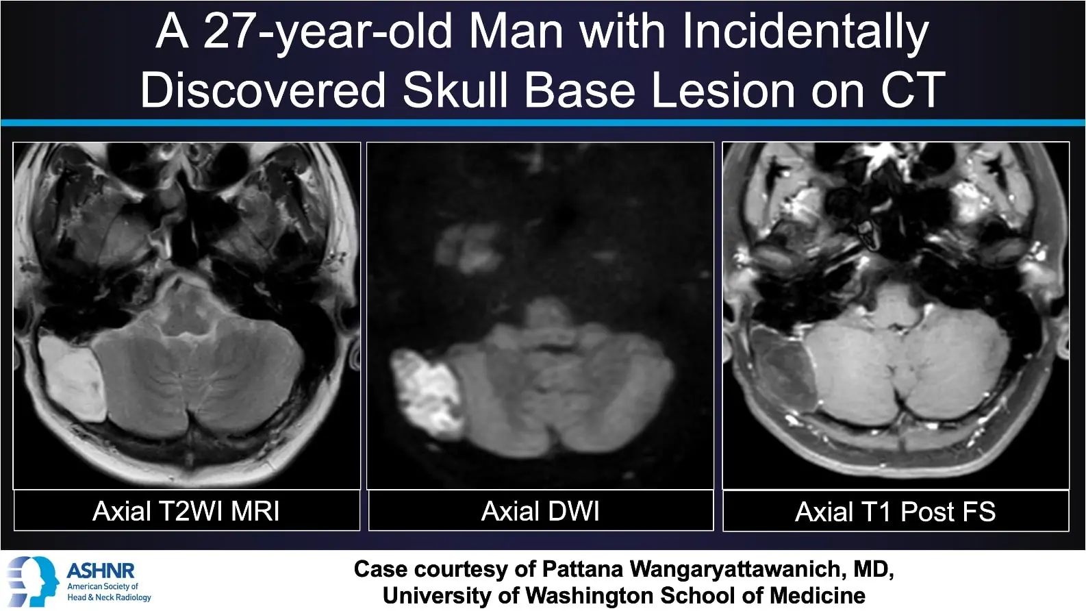
This case features a 27-year-old man with an incidentally discovered skull base lesion on CT, further evaluated with MRI and diagnosed as an intradiploic epidermoid cyst.
Imaging overview:
Axial T2-weighted MRI shows a sharply marginated, expansile lesion within the skull base diploic space that is markedly hyperintense relative to brain parenchyma. The lesion demonstrates bright signal and restricted diffusion on DWI, consistent with keratinous content. Post-contrast axial T1 fat-suppressed images show no internal enhancement, confirming its nonvascular, cystic nature.
Clinical insight:
Intradiploic epidermoid cysts are benign, slow-growing lesions arising from ectopic ectodermal cells trapped within the calvarial diploic space. They often present as painless swellings, headaches, or incidental findings in young adults. Recognizing the combination of T1 hypointensity, T2 hyperintensity, diffusion restriction, and lack of enhancement helps distinguish epidermoid cysts from other skull base masses and supports appropriate neurosurgical referral for resection.
Case courtesy of Pattana Wangaryattawanich, MD
University of Washington School of Medicine